Macrostomum Lignano was discovered in 1995 on a beach bordering the city of Lignano (Italy). Its natural habitat is the high-tide interstitial sand fauna of the Northern Adriatic Sea beaches.
With the support of the Eugene Berezikov team (ERIBA, Netherlands), Macrostomum cultures (NL10 strain) have been introduced in the lab in 2016.
Macrostomum lignano (wikipedia) is an emerging invertebrate model that possesses many advantages over the classical worm models such as Caenorhabditis elegans (nematodes), Schmidtea mediterranea or Dugesia japonica (Planaria). In particular, this platyhelminth is extensively studied for its regeneration properties in order to address fundamental questions regarding stem cell biology, regeneration, and aging.
All developmental stages are also quite easy to observe. Macrostomum is transparent and most organs can be observed macroscopically with a simple inverted binocular microscope. Appropriate staining of entire worms allows observation of deep structures and cellular organization of organs using confocal microscopy (Fig. 1).

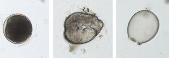
M. lignano is a hermaphrodite worm but does not carry out self-fertilization. Adults lay single cell eggs of about 100 μm in diameter that can be injected thus allowing genetic experiments. Embryonic development takes about 5 days until eggs hatch from the eggshell (Fig. 2). The generation time is about 18 days.
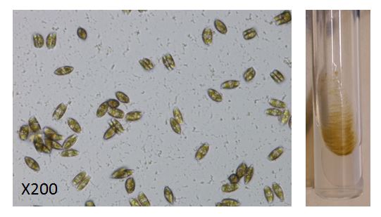
M. lignano is easily maintained in the laboratory using reconstituted seawater in a phyto-incubator at 20°C with a 14 h/10 hour day/night cycle. Worms exclusively feed on the diatom Nitzschia curvilineata (Fig. 3).